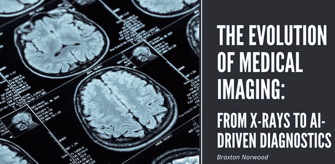
Medical imaging has made significant advancements over the past century, revolutionizing diagnostics and treatment.
The discovery of X-rays in 1895 by Wilhelm Conrad Roentgen marked a breakthrough, enabling the first non-invasive images of the human body. While X-rays are excellent for imaging bones, they fall short with soft tissues, which lead to the development of other techniques.
Ultrasound, based on sonar technology from World War II, is widely used for soft tissue imaging, such as prenatal exams. It’s affordable, portable, and free of radiation, unlike X-rays.
In the 1970s, Computed Tomography (CT) was introduced, allowing detailed cross-sectional images of the body using X-rays. This technology, developed by Sir Godfrey Hounsfield and Allan Cormack, expanded diagnostic capabilities significantly.
Magnetic Resonance Imaging (MRI), rooted in nuclear magnetic resonance principles, emerged around the same time. MRI uses magnetic fields and radio waves to image soft tissues, offering exceptional detail without radiation exposure.
Unlike the previous techniques, which are anatomical, Positron Emission Tomography (PET) provides functional imaging, allowing doctors to observe and measure metabolic processes in the body, crucial for early disease detection and monitoring. PET is often used in tandem with CT or MRI to correlate function with structure.
Today, Artificial Intelligence (AI) is transforming medical imaging by improving diagnostic accuracy and efficiency, analyzing vast datasets to detect patterns that humans might miss, thus advancing personalized treatment.

